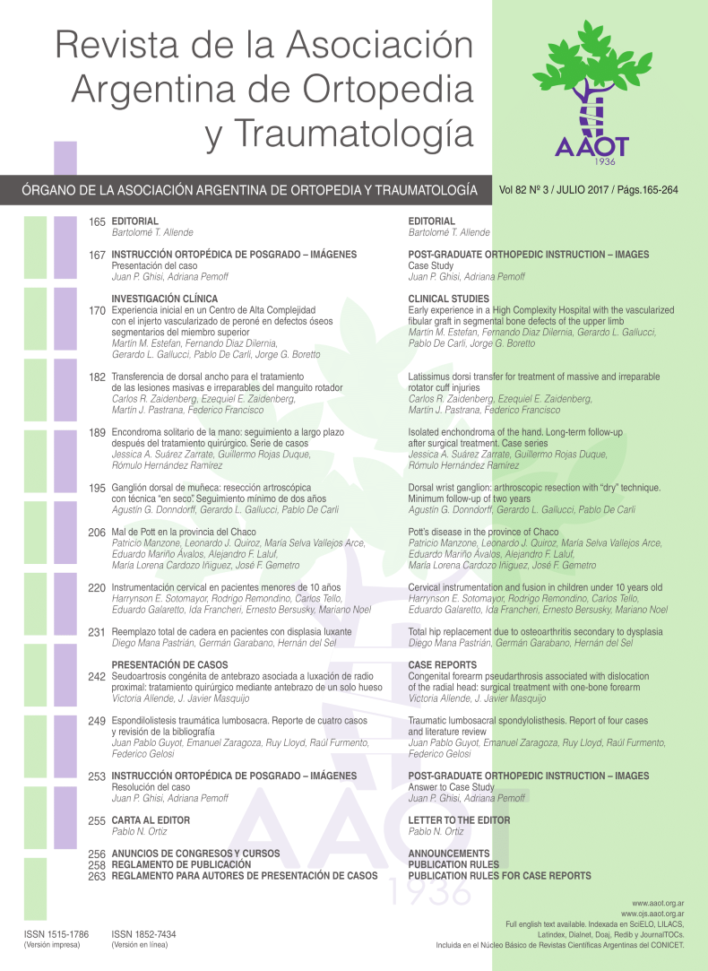
Cervical Instrumentation and Fusion in Children Under 10 Years Old
Abstract
Objective:To assess the postoperative results of cervical instrumented fusion in children under 10 years old, description of the surgical technique and complications.Introduction: The pediatric cervical spine implants and construction designs had rapidly evolved in the last decade. Anatomic and biomechanical differences between pediatric and adult cervical spine required a thorough preoperative planning because this implants were originally designed for adults.Methods:Retrospective descriptive study. Level of Evidence: IV. We evaluated 28 children with different cervical pathology treated with instrumented fusion. We performed Roy-Camille’s technique and Magerl’s technique for lateral masses and Goel-Harms’ for C1-C2 fusion, and occipital-cervical fusion with occipital plates or screws. Inclusion criteria were: children under 10 years, cervical spine deformity or instability, no prior history of spine surgery, minimum follow-up of 2 years.Results:We reported 17 girls and 11 boys with a mean age of 6yr 1mo (1yr 6mo to 10yr). Mean follow-up was 3yr 8mo (2yr to 10yr). The overall complications rate was 32.1%. The highest complication rates were associated to: genetic syndrome, 6 cases (21.4%); skeletal dysplasia, 3 cases (10.7%). Thirteen patients were younger than 6 years, 7 of them had complications (53.8%).Conclusion:Cervical instrumented fusion in patients younger than 10 years old proved to be a feasible and safe surgical procedure. Vertebral dysplasia, congenital, syndromic anomalies, and being younger than 6 years old are the main factors associated with high complication rates.Downloads
References
Harms J., Melcher RP. Posterior C1–C2 fusion with polyaxial screw and rod fixation. Spine 26:2467–2471
Leonard J, Wright N (2006) Pediatric atlantoaxial fixation with bilateral, crossing C-2 translaminar screws. Technical note. J Neurosurg 104:59–63
Jea A, Taylor, Dirks PB, Kulkarni AV, Rutka JT, Drake JM. Incorporation of C-1 lateral mass screws in occipitocervical and atlantoaxial fusions for children 8 years of age or younger. Technical note. J Neurosurg 107:178–183
Schmidt, R., Koller H., Wilke H., Brade J., Zenner J., Meier O. M. The Impact of Cervical Pedicle Screws for Primary Stability in Multilevel Posterior Cervical Stabilizations. Spine 2010;35:E1167–E1171
Dickman CA, Sonntag VK, Papadopoulos SM, Hadley MN (1991) The interspinous method of posterior atlantoaxial arthrodesis. J Neurosurg 74:190–198.
Gallie WE (1937) Skeletal traction in the treatment of fractures and dislocations of the cervical spine. Ann Surg 106:770–776
Hedequist D., Hresko T., Proctor M. Modern Cervical Spine Instrumentation in Children Spine.2008;33:379–383
Prakash S., William G. M., S. A. Sah., Mihir T., Colleen D., Laurens H., J. Campbell, MD. Occipitocervical Fusion in Skeletal Dysplasia. Spine 2014;39:E912–E918.
Michael C. Ain, MD, Kaisorn L. Chaichana, BS, and Joshua G. Schkrohowsky, BS. Retrospective Study of Cervical Arthrodesis in Patients With Various Types of Skeletal Dysplasia. Spine 2006;31:E169–E174
Ryoji Tauchi • Shiro Imagama • Zenya Ito • Kei Ando • Kenichi Hirano • Akio Muramoto. Complications and outcomes of posterior fusion in children with atlantoaxial instability. Eur Spine J (2012) 21:1346–1352
Doyle, J. Scott MD; Lauerman, William C. MD; Wood, Kirkham B. MD; Krause, David R. Complications and Long-Term Outcome of Upper Cervical Spine Arthrodesis in Patients With Down Syndrome. Spine Volume 21(10), 15 May 1996, p 1223–1231
Rishimugesh Kanna P, Ajoy Prasad Shetty, Rajasekaran S. Anatomical Feasibility of Pediatric Cervical Pedicle Screw Insertion by Computed Tomographic Morphometric Evaluation of 376 Pediatric Cervical Pedicles. Spine 2011 ; 36 : 1297 – 1304
Ferri-de-Barros F., Little D. G., Bridge C., Cummine J, Cree K. A. Atlantoaxial and Craniocervical Arthrodesis in Children. A Tomographic Study Comparing Suitability of C2 Pedicles and C2 Laminae for Screw Fixation. Spine 2010;35:291–293
Abdullah K.G., Steinmetz M. P., Mroz T. E. Morphometric and Volumetric Analysis of the Lateral Masses of the Lower Cervical Spine. Spine 2009;34:1476–1479
Jeanneret B, Magerl F (1992) Primary posterior fusion C1/2 in odontoid fractures: indications, technique, and results of transarticular screw fixation. J Spinal Disord 5:464–475
Roy-Camille R., Saillant G., Laville C. Treatment of lower cervical spinal injuries—C3 to C7. Spine 1992. 17(10 Suppl):S442–S446
Heller JG, Carlson GD, Abitbol JJ. Anatomic comparison of the Roy-Camille and Magerl techniques for screw placement in the lower cervical spine. Spine 1991. 16(10 Suppl):S552–S557
Grob D, Crisco JJ III, Panjabi MM, et al. Biomechanical evaluation of four different posterior atlantoaxial fixation techniques. Spine 1992;17:480–90.
Brockmeyer DL, York JE, Apfelbaum RI (2000) Anatomical suitability of C1–2 transarticular screw placement in pediatric patients. J Neurosurg 92:7–11
Heuer GG, Hardesty DA, Bhowmick DA, Bailey R, Magge SN, Storm PB (2009) Treatment of pediatric atlantoaxial instabilitywith traditional and modified Goel-Harms fusion constructs. Eur. Spine J. 2009, 18:884–892
- Artrodesis cervical pedriatrica (Español (España))
- Figura 9. Imágenes Intraoperatorias: Decompresión posterior más fusión posterior instrumentada occipito torácica (Español (España))
- Figura 10. Imágenes Postoperatorio inmediata: fusión occipito torácica más halo chaleco (Español (España))
- Figura 11. Imágenes Radiográficas 2 años postoperatoria (Español (España))
- Figura 12. Tomografía reconstrucción 3 D. 2 años postoperatorio (Español (España))
- manuscrito final 15 julio (Español (España))
- Figura 8. Radiografías cervical AP y Lateral (Español (España))
- Figura 13. Cifosis cervical asociada a compresión medular (Español (España))
- caso clinico 2 (Español (España))
- Figura 8 A. Resonancia Magnetica. Compresión medular severa y mielomalacia asociada. (Español (España))
- Figura 14. 4 meses postoperatorio. Fusión posterior instrumentada occipito - torácico (Español (España))
- Figura 15. 11 meses postoperatorio. Pseudoartrosis occipito -C1. sin deterioro del status neurológico (Español (España))
- Figura 2. Tomografia Axial. Inestabilidad Atlanto - Axial (Español (España))
- Figura 3. Resonancia Magnética sin evidencia de lesión medular (Español (España))
- Figura 4. Tomografía reconstrucción 2D. Muesta inestabilidad C1-C2. Hipoplasia de Odontoides (Español (España))
- Figura 5. Radiografias post. caída, muestra full out de tornillos occipitales (Español (España))
- Figura 6. Tomografia reconstrucción 2 D. Full out de tornillos occipitales (Español (España))
- Figura 7. Radiografías Post revisión AP y Lateral (Español (España))
- resumen en ingles (Español (España))
- Figura 1. Radiografias preoperatorias AP y Laterales (Español (España))
Copyright (c) 2017 Revista de la Asociación Argentina de Ortopedia y Traumatología

This work is licensed under a Creative Commons Attribution-NonCommercial-ShareAlike 4.0 International License.
Manuscript acceptance by the Journal implies the simultaneous non-submission to any other journal or publishing house. The RAAOT is under the Licencia Creative Commnos Atribución-NoComercial-Compartir Obras Derivadas Igual 4.0 Internacional (CC-BY-NC.SA 4.0) (http://creativecommons.org/licences/by-nc-sa/4.0/deed.es). Articles can be shared, copied, distributed, modified, altered, transformed into a derivative work, executed and publicly communicated, provided a) the authors and the original publication (Journal, Publisher and URL) are mentioned, b) they are not used for commercial purposes, c) the same terms of the license are maintained.
In the event that the manuscript is approved for its next publication, the authors retain the copyright and will assign to the journal the rights of publication, edition, reproduction, distribution, exhibition and communication at a national and international level in the different databases. data, repositories and portals.
It is hereby stated that the mentioned manuscript has not been published and that it is not being printed in any other national or foreign journal.
The authors hereby accept the necessary modifications, suggested by the reviewers, in order to adapt the manuscript to the style and publication rules of this Journal.