Factors associated with severe pain in patients with plantar fasciitis: An association analysis
Main Article Content
Abstract
Materials and Methods: We conducted a prospective study in a cohort of patients with PF diagnosis. Each participant completed an ordinal pain scale (1-10) for first-step pain and end-of-day pain, and Foot Function Index-Revised (FFI-R) surveys at enrollment. Also, patient demographics were evaluated. The ankle joint dorsiflexion, the range of motion (ROM) for the first metatarsophalangeal joint (MTPJ), the gastrocnemius tightness, and the popliteal angle were evaluated through standard tests.
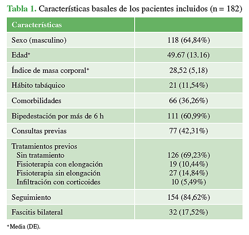
Results: Our study included 214 participants, of which 64% (118 patients) were males, the average age was 49.67 years (SD 13.16) and the average BMI was 28.53 (SD 5.18). The multivariate analysis showed that the risk of having a Visual Analog Scale (VAS) score ≥8 increased when the patient reported standing for more than 6 hours (OR=1.17; P=0.03; CI95%: 1.02-1.359). The risk of a >8-VAS score was higher when the level of ankle dorsiflexion was <0 (OR=1.20; P=0.03; CI95%: 1.02-1.41).
Conclusion: Our findings indirectly support the hypothesis that limited ankle dorsiflexion ROM plays a role as a risk factor associated with VAS scores ≥8, in PF patients.
Level of Evidence: IV
Downloads
Metrics
Article Details
Manuscript acceptance by the Journal implies the simultaneous non-submission to any other journal or publishing house. The RAAOT is under the Licencia Creative Commnos Atribución-NoComercial-Compartir Obras Derivadas Igual 4.0 Internacional (CC-BY-NC.SA 4.0) (http://creativecommons.org/licences/by-nc-sa/4.0/deed.es). Articles can be shared, copied, distributed, modified, altered, transformed into a derivative work, executed and publicly communicated, provided a) the authors and the original publication (Journal, Publisher and URL) are mentioned, b) they are not used for commercial purposes, c) the same terms of the license are maintained.
In the event that the manuscript is approved for its next publication, the authors retain the copyright and will assign to the journal the rights of publication, edition, reproduction, distribution, exhibition and communication at a national and international level in the different databases. data, repositories and portals.
It is hereby stated that the mentioned manuscript has not been published and that it is not being printed in any other national or foreign journal.
The authors hereby accept the necessary modifications, suggested by the reviewers, in order to adapt the manuscript to the style and publication rules of this Journal.
References
2. Tisdel CL, Donley BG, Sferra JJ. Diagnosing and treating plantar fasciitis: a 511 conservative approach to plantar heel pain. Cleve Clin J Med 1999;66(4):231-5. https://doi.org/10.3949/ccjm.66.4.231
3. Irving DB, Cook JL, Young MA, Menz HB. Impact of chronic plantar heel pain 524 on health-related quality of life. J Am Podiatr Med Assoc 2008;98(4):283-9. https://doi.org/10.7547/0980283
4. Arslan A, Koca TT, Utkan A, Sevimli R, Akel I. Treatment of chronic plantar heel pain 310 with radiofrequency
neural ablation of the first branch of the lateral plantar nerve and 311 medial calcaneal nerve branches. J Foot Ankle Surg 2016;55:767-71. https://doi.org/10.1053/j.jfas.2016.03.009
5. Orchard J. Plantar fasciitis-Clinical review. BMJ 2012;345:e6603. https://doi.org/10.1136/bmj.e6603
6. Bennett PJ, Patterson C, Wearing S, Baglioni T. Development and validation of a questionnaire designed to measure foot-health status. J Am Podiatr Med Assoc 1998;88(9):419-28.
https://doi.org/10.7547/87507315-88-9-419
7. Reips UD, Funke F. Interval-level measurement with visual analogue scales in Internet-based research: VAS
Generator. Behavior Research Methods 2008;40(3):699-704. https://doi.org/10.3758/BRM.40.3.699
8. Coughlin MJ, Shurnas PS. Hallux rigidus: demographics, etiology, and radiographic assessment. Foot Ankle Int
2003;24:731-43. https://doi.org/10.1177/107110070302401002
9. Silverfskiold N. Reduction of the uncrossed two-joints muscles of the leg to one-joint muscles in spastic conditions. Acta Chir Scand 1924;56:315-28.
10. Quintana E, Albuquerque F. Evidencia científica de los métodos de evaluación de la elasticidad de la musculatura isquiotibial. Osteopat Cient 2008;3:115-24. https://doi.org/10.1016/S1886-9297(08)75760-6
11. Riddle DL, Pulisic M, Pidcoe P, Johnson RE. Risk factors for plantar fasciitis: a matched case-control study. J Bone Joint Surg Am 2003;85(5):872-7. https://doi.org/10.2106/00004623-200305000-00015
12. Aranda Y, Munuera PV. Plantar fasciitis and its relationship with hallux limitus. J Am Podiatr Med Assoc
2014;104(3):263-8. https://doi.org/10.7547/0003-0538-104.3.263
13. Amis J, Jennings L, Graham D, Graham CE. Painful heel syndrome: radiographic and treatment assessment. Foot Ankle 1988;9(2):91-5. https://doi.org/10.1177/107110078800900206
14. Taunton JE, Ryan MB, Clement DB, McKenzie DC, Lloyd-Smith DR. Plantar fasciitis: a retrospective analysis of
267 cases. Phys Ther Sport 2002;3:57-65. https://doi.org/10.1054/ptsp.2001.0082
15. Rome K, Howe T, Haslock I. Risk factors associated with the development of plantar heel pain in athletes. Foot
2001;11:119-25. https://doi.org/10.1054/foot.2001.0698
16. Cole C, Seto C, Gazewood J. Plantar fasciitis: evidence-based review of diagnosis and therapy. Am Fam Physician 2005;72(11):2237-42. PMID: 16342847
17. Landorf KB, Menz HB. Plantar heel pain and fasciitis. BMJ Clin Evid 2008. pii: 1111. PMID: 19450330
18. Sweeting D, Parish B, Hooper L, Chester R. The effectiveness of manual stretching in the treatment of plantar heel pain: a systematic review. J Foot Ankle Res 2011;4:19. https://doi.org/10.1186/1757-1146-4-19
19. Bolívar YA, Munuera PV, Padillo JP. Relationship between tightness of the posterior muscles of the lower limb and plantar fasciitis. Foot Ankle Int 2013;34(1):42-8. https://doi.org/10.1177/1071100712459173
20. Budiman-Mak E, Conrad K, Stuck R, Matters M. Theoretical model and Rasch analysis to develop a revised Foot Function Index. Foot Ankle Int 2006;27(7):519-27. https://doi.org/10.1177/107110070602700707

