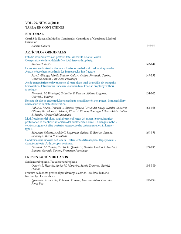
Changes in the cervical alignment after posterior transpedicular instrumentation in Lenke type 1 adolescent idiopathic scoliosis
Main Article Content
Abstract
Downloads
Metrics
Article Details

This work is licensed under a Creative Commons Attribution-NonCommercial-ShareAlike 4.0 International License.
Manuscript acceptance by the Journal implies the simultaneous non-submission to any other journal or publishing house. The RAAOT is under the Licencia Creative Commnos Atribución-NoComercial-Compartir Obras Derivadas Igual 4.0 Internacional (CC-BY-NC.SA 4.0) (http://creativecommons.org/licences/by-nc-sa/4.0/deed.es). Articles can be shared, copied, distributed, modified, altered, transformed into a derivative work, executed and publicly communicated, provided a) the authors and the original publication (Journal, Publisher and URL) are mentioned, b) they are not used for commercial purposes, c) the same terms of the license are maintained.
In the event that the manuscript is approved for its next publication, the authors retain the copyright and will assign to the journal the rights of publication, edition, reproduction, distribution, exhibition and communication at a national and international level in the different databases. data, repositories and portals.
It is hereby stated that the mentioned manuscript has not been published and that it is not being printed in any other national or foreign journal.
The authors hereby accept the necessary modifications, suggested by the reviewers, in order to adapt the manuscript to the style and publication rules of this Journal.
References
cervical spine in compression is increased under a follower load. Spine 2000;25:1548-54.
2. Panjabi MM. Cervical spine models for biomechanical research. Spine 1998;23:2684-700.
3. Harrison DD, Harrison DE, Janik TJ, Cailliet R, Ferrantelli JR, Holland B, et al. Modeling of the sagittal cervical spine as a
method of discriminate hypolordosis. Results of elliptical and circular modeling in 72 asymptomatic subjects, 52 acute neck pain
and 70 chronic neck pain subjects. Spine 2004;29(22):2485-92.
4. Reilly CW, Mulpuri K, Perdios A. Evidence-based medicine analysis of all pedicle screw constructs in AIS. Spine
2007;32(19S):109-14.
5. Kim YJ, Lenke LG, Kim J, Bridwell KH, Cho SK, Chen G, et al. Comparative analysis of pedicle screw vs. hybrid
instrumentation in posterior spinal fusion of AIS. Spine 2006;31:291-8.
6. Watanabe K, Lenke LG, Bridwell KH, Kim YJ, Watanabe K, Kim YW, et al. Comparison of radiographic outcomes for the
treatment of scoliotic curves greater than 100 degrees: wires vs. hooks vs. screws. Spine 2008;33:1084-92.
7. Suk SI, Kim WJ, Kim JH, Lee SM. Restoration of thoracic kyphosis in the hypokyphotic spine: a comparison between
multiple-hook and segmental pedicle screw fixation in AIS. J Spinal Disord 1999;12:489-95.
8. Suk SI, Lee SM, Chung ER, Kim JH, Kim WJ, Sohn HM. Determination of distal segmental pedicle screw fixation in single
thoracic idiopathic scoliosis. Spine 2003;28:484-91.
9. Lowenstein JE, Matsumoto H, Vitale MG, Weidenbaum M, Gomez JA, Young-In Lee FYI, et al. Coronal and sagittal plane
correction in AIS: comparison between all pedicle screws vs. hybrid thoracic hook lumbar screws constructs. Spine 2007;32:448-
52.
10. Sucato DJ, Agrawal S, O´Brien MF, Lowe TG, Richards SB, Lenke L. Restoration of thoracic kyphosis after operative
treatment of AIS. A multicenter comparison of three surgical approaches. Spine 2008;30(24):2630-6.
11. Harms Study Group. Preservation of thoracic kyphosis is critical to maintain lumbar lordosis in the surgical treatment of AIS.
Spine 2010;35(14):1365-70.
12. Lee SM, Suk SI, Chung ER. Direct vertebral rotation: a new technique of three-dimensional deformity correction with
segmental pedicle screw fixation in AIS. Spine 2004;29:343-9.
13. Gerald M, Gibson MJ, Quan Y. Correction of main thoracic adolescent idiopathic scoliosis using pedicle screws
instrumentation. Does higher implant density improve correction? Spine 2010;35:562-7.
14. Hillibrand AS, Tannenbaum DA, Graziano GP, Loder RT, Hensinger RN. The sagittal aligment of the cervical spine in
adolescent idiopathic scoliosis. J Pediatr Orthop 1995;15:627-32.
15. Clement JL, Chau E, Kimkpe C, Vallade MJ. Restoration of thoracic kyphosis by posterior instrumentation in AIS.
Comparative radiographic analysis of two methods of reduction. Spine 2008;33(14):1579-87.
16. Westrick ER, Ward TW. AIS: 5-year to 20-year evidence-based surgical results. J Pediatr Orthop 2011;31:s61-68.
17. Harms Study Group. Evaluation of proximal junctional kyphosis in AIS following pedicle screw, hook, or hybrid
instrumentation. Spine 2010;35(2):177-81.
18. Edgar MA, Metha MH. Long term follow up of fused and unfused idiophatic scoliosis. J Bone Joint Surg Br 1988;70:712-6.
19. Moskowitz A, Moe JH, Winter RB, Binner H. Long term follow up of scoliosis fusion. J Bone Joint Surg Am 1980;62:364-76.
20. Canavese F, Turcot K, De Rosa V, de Coulon G, Kaelin A. Cervical spine sagittal aligment variations following posterior spinal
fusion and instrumentation for AIS. Eur Spine J 2011;20:1141-8.

