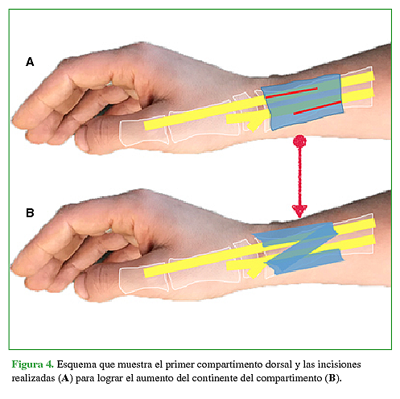
De Quervain tenosynovitis: New enlargement plasty of the first dorsal compartment Anatomical study and clinical retrospective study
Main Article Content
Abstract
Materials and Methods: Anatomical study of 12 cadaver wrists to corroborate first compartment enlargement and its relationship to the sensitive branch of the radial nerve. Clinical retrospective study of patients who had undergone surgery between 2014 and 2019 due to De Quervain tenosynovitis (DQT)refractory to nonsurgical management, over 18 years of age, with no previous surgical history and a 12-month minimum follow-up. The 22-patient series was divided into two groups: enlargement (Group A) and simple release (Group B). Average ages were 47 years (Group A) and 50 years (Group B). Subjective outcome was evaluated by the visual analogue scale (VAS) for pain, the Quick-DASH score, and the Patient Satisfaction Questionnaire Short Form (PSQ-18). Objective outcome was evaluated by goniometry and dynamometry tests.
Results: The anatomical study proved the increase of the FDC laxity and its close relationship to thesensitive branch of the radial nerve. The clinical study follow-up periods were of 24 months in Group A and 50 months in Groups B. Average VAS scores were 0.5/10 in Group A and 1/10 in Group B). Satisfaction index was 97% in both groups. Quick-DASH scores, and goniometry and dynamometry tests yielded no significant differences.
Conclusions: The new enlargement plasty of the FDC for the surgical treatment of DQT in this anatomical and clinical study proved to be a reproducible and effective technique.
Key words: De Quervain; enlargement; sensitive branch of the radial nerve.Level of Evidence: III
Downloads
Metrics
Article Details
Manuscript acceptance by the Journal implies the simultaneous non-submission to any other journal or publishing house. The RAAOT is under the Licencia Creative Commnos Atribución-NoComercial-Compartir Obras Derivadas Igual 4.0 Internacional (CC-BY-NC.SA 4.0) (http://creativecommons.org/licences/by-nc-sa/4.0/deed.es). Articles can be shared, copied, distributed, modified, altered, transformed into a derivative work, executed and publicly communicated, provided a) the authors and the original publication (Journal, Publisher and URL) are mentioned, b) they are not used for commercial purposes, c) the same terms of the license are maintained.
In the event that the manuscript is approved for its next publication, the authors retain the copyright and will assign to the journal the rights of publication, edition, reproduction, distribution, exhibition and communication at a national and international level in the different databases. data, repositories and portals.
It is hereby stated that the mentioned manuscript has not been published and that it is not being printed in any other national or foreign journal.
The authors hereby accept the necessary modifications, suggested by the reviewers, in order to adapt the manuscript to the style and publication rules of this Journal.
References
2. Moore JS. De Quervain’s tenosynovitis: Stenosing tenosynovitis of the first dorsal compartment. J Occup Environ Med 1997;39:990-1002. https://doi.org/10.1097/00043764-199710000-00011
3. Altay M, Erturk C, Isikan U. De Quervain’s disease treatment using partial resection of the extensor retinaculum: A short-term results survey. Orthop Traumatol Surg Res 2011;97(5):489-93. https://doi.org/10.1016/j.otsr.2011.03.015
4. Ramesh R, Britton JM. A retinacular sling for subluxing tendons of the first extensor compartment. A case report. J Bone Joint Surg Br 2000;82(3):424-5. https://doi.org/10.1302/0301-620x.82b3.9867
5. Perno-Ioanna D, Papaloïzos M. A comprehensive approach including a new enlargement technique to prevent
complications after De Quervain tendinopathy surgery. Hand Surg Rehabil 2016;35(3):183-9.
https://doi.org/10.1016/j.hansur.2016.03.002
6. Rogozinski B, Lourie G. Dissatisfaction after first dorsal compartment release for De Quervain tendinopathy.
J Hand Surg Am 2016;41:117-9. https://doi.org/10.1016/j.jhsa.2015.09.003
7. Abrams R, Brown R, Botte M. The superficial branch of the radial nerve: An anatomic study with surgical
implications. J Hand Surg Am 1992;17(6):1037-41. https://doi.org/10.1016/s0363-5023(09)91056-5
8. Kilic A, Kale A, Usta A, Bilgili F, Yabukcuoglu Y, Sökücü S. Anatomic course of the superficial branch of the
radial nerve in the wrist and its location in relation to wrist arthroscopy portals: A cadaveric study. Arthroscopy
2009;25(11):1261-4. https://doi.org/10.1016/j.arthro.2009.05.015
9. Jordaan P, Kang Wang C, Yew Ng C. Management of painful cutaneous neuromas around the wrist. Orthop Trauma 2017;30:1-6. https://doi.org/10.1016/j.mporth.2017.05.006

